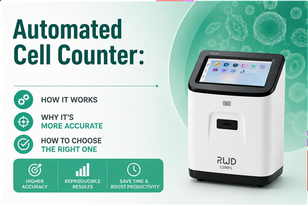
Automated Cell Counter: How It Works, Why It’s More Accurate, and How to Choose the Right One
Cell counting is one of the most frequently performed procedures in cell biology laboratories. Yet many researchers still rely on manual hemocytometer counting—a method that can be time-consuming, operator-dependent, and difficult to reproduce.
Automated cell counters were developed to solve these challenges by providing rapid, standardized, and highly reproducible cell analysis.
Table of Content
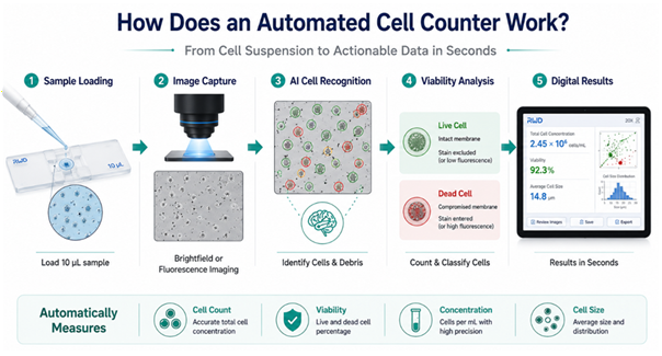
How Does an Automated Cell Counter Work?
The process begins when:
Eg:
Step |
Process |
| 1 | Load sample |
| 2 | Capture image |
| 3 | Identify cells |
| 4 | Calculate results |
| 5 | Export data |
Which Type of Cell Counter Do You Need?
The market typically offers three categories: brightfield counters, fluorescent counters, and Brightfield + Fluorescence Hybrid Systems.
Type |
Features and Applications |
| Brightfield Counters | Standard for routine cell line maintenance; most commonly used with trypan blue staining for cell viability assessment. |
| Fluorescent Cell Counters | Incorporate multiple light channels; suitable for complex samples such as Peripheral Blood Mononuclear Cells (PBMCs) or stem cells; can detect GFP or RFP expression to evaluate transfection efficiency alongside standard cell counts. |
| Brightfield + Fluorescence Hybrid Systems | Support fluorescence channel expansion for advanced workflows; for example, the C100/C100-SE Automated Cell Counter supports modular fluorescence cube integration. |
Why is an Automated Cell Counter More Accurate?
Manual counting may introduce error sources, including:
- Uneven sample mixing
- Inconsistent chamber loading
- Subjective cell identification
- Counting fatigue
- Operator-to-operator interpretation differences
Automated systems improve cell counting accuracy through standardization:
- Consistent imaging parameters
- Algorithm-based thresholding
- Repeatable field analysis
The increased accuracy is essential for several core applications. These include tumor research for passaging cell lines and pre-detection of apoptosis, immunology for counting Peripheral Blood Mononuclear Cells (PBMCs) and lymphocytes, cell biology research for primary cells, and cell quality control for stem cells or Cytokine-Induced Killer (CIK) cells. Fluorescent models are specifically required for detecting transfection efficiency in gene expression studies.
The key differences are summarized below:
Dimension |
Manual Counting |
Automated Counting |
| Operation | Operator dependent | More standardized workflow |
| Cell identification | Subjective judgment | Algorithm-assisted recognition |
| Reproducibility & consistency | Variable between users and runs | More consistent results |
| Throughput | Relatively low | Higher throughput |
| Fatigue impact | May affect accuracy over time | Reduces fatigue-related variation |
| Data recording | Manual entry, prone to errors | Digital recording and storage |
How to Use an Automated Cell Counter
The workflow of an automated cell counter is designed to simplify and standardize cell analysis, typically involving four key steps:
Step 1: Sample Preparation
Prepare a uniform cell suspension by dissociating cells from the culture vessel (e.g., using Trypsin) and resuspending them in a known volume of buffer or media. Ensure the sample is well mixed and free of large clumps to support accurate analysis.
Step 2: Staining
For brightfield counting, mix the sample with trypan blue (commonly 1:1) to assess viability.
For fluorescence-based counting, add appropriate dyes (e.g., AO/PI) to distinguish live and dead cells based on DNA fluorescence.
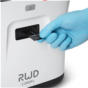
Step 3: Load & Analyze
Load the prepared sample into a disposable counting slide and insert it into the instrument. The system automatically captures images, identifies cells, and calculates parameters such as cell concentration, size, and viability.
Step 4: Review Results
Results are displayed on the screen for review. Users can verify cell recognition through images and export data for further analysis or record keeping.
How to Choose an Automated Cell Counter?
Selecting the appropriate counter depends on the needs of the laboratory, including the cell types being studied and the daily volume of samples processed.
1. Accuracy Requirements
If a laboratory focuses on standard cell lines, a brightfield counter with a CV of less than 5% is usually sufficient. Accuracy requirements are driven by the sensitivity of downstream experiments. High-stakes research, such as pharmaceutical screening, demands the highest level of precision.
2. Throughput Requirements
Throughput refers to how many samples a lab processes. For most academic labs, a single-slide counter that provides results in 10 seconds is adequate.
However, labs processing hundreds of samples daily might look for units that support multi-chamber slides or have faster processing algorithms to reduce “wait time” between samples.
3. Fluorescence Capability
The decision to include fluorescence depends on the sample complexity. If you are working with primary cells, tissue digests, or blood, a fluorescent counter is necessary to distinguish cells from red blood cells or debris.
It also allows for the assessment of transfection efficiency by measuring fluorescent protein expression.
4. Data Management Capability
Data management is another critical factor. Ensure the device supports the export formats your lab uses and offers massive historical data storage capacity.
Pro Tips: For small and medium-sized laboratories or university teaching labs, prioritize models that are cost-effective, easy to operate, and support common cell types.
Large research institutions or core facilities should prioritize higher throughput, expanded fluorescence channels, and larger storage capacity to meet different research needs.
Quick Selection Checklist
Before purchasing, consider the following:
- Are your samples standard cell lines, or primary cells / heterogeneous samples?
- Do you require fluorescence capability (e.g., AO/PI staining or GFP/RFP expression analysis)?
- What level of accuracy and reproducibility is required (e.g., acceptable CV range)?
- What is your laboratory’s daily sample throughput?
- Is cell viability analysis or other functional assays required?
- Do your samples contain significant debris, red blood cells, or cell aggregates?
- What are your data management requirements (e.g., export formats, storage capacity, traceability)?
- Does the system need to be compatible with existing workflows or consumables?
Recommended Solutions for Different Research Needs
Based on different research needs, the following solutions are commonly recommended:
Application |
Recommended System |
| Routine culture | C100-SE |
| Cell viability | C100 |
| Primary cells | C200FL |
| Transfection studies | C200FL |
C100/C100-SE Automated Cell Counters
Designed for efficient brightfield counting, with the C100 additionally equipped with a fluorescence cube. These counters can accurately distinguish clustered cells, delivering results in approximately 9 seconds.
C200FL Fluorescent Cell Counter
For more complex workflows, this counter integrates brightfield and multi-channel fluorescence detection. Features include a 10.1-inch high-definition touchscreen, 6-channel automatic sampling, a smart app interface, and multiple export formats. Counting results are available in just 5 seconds.
BPLabline also supplies high-quality disposable cell counting slides with consistent chamber depth and two independent counting pools. Alongside our products, we provide technical support, training, and guidance to ensure smooth laboratory operations.
With local warehousing in North America, we can respond quickly to urgent requests, while transparent pricing and direct procurement reduce administrative delays and simplify budgeting.
Conclusion
Choosing the right automated cell counter depends on your workflow, sample type, throughput requirements, and data quality expectations.
For routine cell culture, a brightfield counter may be sufficient. For primary cells, stem cells, PBMCs, or transfection studies, fluorescence-enabled systems often provide greater accuracy and confidence.
Understanding these differences helps researchers select a solution that supports both current experiments and future workflow expansion. Explore BPLabline Cell Analysis Solutions →Cell & Molecular Analysis – BP LabLine.