Imaging Techniques for Oral and Dental Applications
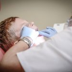
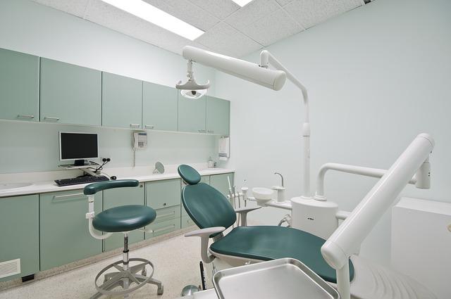
Dental professionals use imaging techniques to diagnose and treat oral health conditions. There are various imaging technologies available, each with its own strengths and weaknesses used by the Dentists. This blog post will discuss the most common dental imaging techniques.
Let’s take a look at them.
Table of Content
Panoramic Radiography
This is a type of dental x-ray that gives your dentist a panoramic or “bird’s-eye” view of all your teeth, as well as your jaws. This radiograph can be used to detect problems with your teeth and jawbone. Panoramic radiography is often used to plan dental implant surgery or orthodontic treatment.
Cone beam CT
Cone beam CT is a relatively new imaging modality that has revolutionized oral and maxillofacial radiology. It provides superior image quality compared to traditional dental radiography and allows for three-dimensional reconstruction of anatomical structures.
CBCT is indicated for numerous oral and maxillofacial applications, including assessment of tooth position, impacted teeth, TMJ disorders, and pathology. The technology is also becoming increasingly popular for implant planning and placement.
Intra-Oral Periapical Radiograph
The intra-oral periapical radiograph is the most commonly used type of dental x-ray. It provides a clear image of the teeth, roots, and surrounding bone. This radiograph is taken with the x-ray film placed inside the mouth. In this technique, the x-ray beam is directed from the back of the mouth towards the teeth.
One advantage of this radiograph is that it can be taken quickly and easily. Additionally, it requires less radiation exposure than other types of dental x-rays. However, because the image is taken from inside the mouth, it can be difficult to get a clear view of all the teeth. This type of radiograph is also not well suited for imaging large mouth areas.
Intraoral bitewing radiograph
The intraoral bitewing radiograph is similar to the intraoral periapical radiograph, but it provides a different view. In this technique, the x-ray film is placed horizontally in the mouth between the teeth. This radiograph is used to detect decay between the teeth and to check the status of existing dental work, such as fillings.
Extraoral bitewing radiograph
The extraoral bitewing radiograph is used to detect cavities between the teeth. This radiograph is taken with the x-ray film placed outside of the mouth. The patient bites down on a small piece of gauze to keep the film in place. It is one of the most common types of dental radiographs.
Panoramic radiograph
The panoramic radiograph provides a broad view of all the teeth in both jaws and is especially useful for viewing the relationships between teeth. It is also helpful in detecting some types of tumors or cysts in the jaw bones. This radiograph is taken with the patient standing or sitting upright and biting on a special plate that helps keep the head still during the imaging process.
Sialography
This radiographic technique is used to visualize the salivary glands. It can help diagnose problems with the salivary glands, such as inflammation or blockages. A small amount of contrast material is injected into the gland so that it can be seen on the x-ray. It is best to avoid eating or drinking for at least an hour before the procedure to minimize the risk of aspiration.
Magnetic resonance imaging (MRI) is another imaging modality that can be used to evaluate salivary glands. This technique does not use ionizing radiation, making it a good choice for pregnant patients or those with other medical concerns that cause exposure to radiation.
The Bottom Line
If you need oral or dental imaging, ask your doctor about CBCT. This technique offers numerous benefits and can help ensure an accurate diagnosis. CBCT is the best option for oral and dental imaging with its low radiation dose and high-quality images. It can provide important information about your condition and help guide treatment decisions.